IB Assessment Statements
IB Biology at Chapel School
sábado, 3 de novembro de 2012
1. Draw and label a diagram of the heart showing the four chambers, associated blood vessels, valves and the route of blood through the heart
The heart is composed by two sides with different functions. The right side receives the blood and pumps it to the lungs and the left side receives the blood and pumps it to the body. It also contains two types of chambers, the atrium and the ventricle, one of each at each side, which makes it 4 chambers (two atria and two ventricles).
The atrioventricular valves connects each atrium to the ventricle below it. The mitral valve connects the left atrium with the left ventricle and the tricuspid valve connects the right atrium with the right ventricle.
The heart connects to blood vessels, being the aorta the largest one, which carries blood away from the heart. Another important vessel is the pulmonary artery, that connects the heart with the lungs. The superior and inferior vena cava are the two largest veins that carry blood into the heart.
2. State that coronary arteries supply heart muscle with oxygen and nutrients
http://click4biology.info/c4b/6/hum6.2.htm#2
Coronary arteries are branches of the aorta and they supply the heart muscle with oxygen and nutrients.
Coronary arteries are branches of the aorta and they supply the heart muscle with oxygen and nutrients.
3. Explain the action of the heart in terms of collecting blood, pumping blood, and opening and closing of valves
http://www.ck12.org/user:kay.teehan@polk-fl.net/concept/Heart-and-Blood-Vessels-%253A%253Aof%253A%253A-MS-Cardiovascular-System/
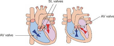
While the right atrium is responsible for collecting blood from the superior vena cava and from the inferior vena cava, the left atrium is responsible for collecting blood from the pulmonary veins. The collected blood then flows into the right and left ventricle and is pumped into the arteries. The atrioventricular and semilunar valves are responsible for controlling and directing the blood flow. When the atria contracts, the atrioventricular valves open and the blood flows through them into the ventricle. The semilunar valves are closed so the ventricle can be filled with blood. Then, the ventricles contract causing a rise in the pressure, that causes the atrioventricular valves to close, preventing back flow of blood into the atria. The semilunar valves then open and allows the blood flow into the arteries, and the atria starts to fill with blood again.When the ventricles stop contracting, it leads to a fall in the pressure and causes the semilunar valves to close, preventing back flow of blood, as well as it allows the atrioventricular valves to re-open, and the cycle repeats.
4. Outline the control of the heartbeat in terms of myogenic muscle contraction, the role of the pacemaker, nerves, the medulla of the brain and epinephrine (adrenaline)
- Myogenic muscle contraction means the heart muscle can contract by itself.
- In the wall of the right atrium, the pacemaker (SA nodes) is located, region that initiates each heart contraction, and whenever the pacemaker sends a signal, the heart contracts and causes a heartbeat.
- Nerves and hormones are responsible for transmitting messages to the pacemaker; the adrenaline, for example carries the massages from the medulla of the brain to the pacemaker telling it to speed up the beating.
- Another nerve sends a massage telling the pacemaker to slow down the beatings.
5. Explain the relationship between the structure and function of arteries, capillaries and veins
http://click4biology.info/c4b/6/hum6.2.htm
Arteries have a thick outer layer of longitudinal collagen and elastic fibers to avoid leaks. It also contains a thick wall to deal with the high pressures, as well as a narrow lumen to maintain these high pressures and thick layers of muscle fibers to help pump the blood after a hearth beat. Its function is to carry blood away from the heart to the body, under high pressure.
Veins are responsible for returning the blood to the heart, under low pressure. It is composed of a thin outer layer of longitudinal collagen and elastic fibers, since there is no risk or leaking and bursting. It also has a thin walls that allows the vein to be pressed by muscles, helping to move the blood. It contains thin layers of muscle fibers, since the blood does not flow in pulses, and the lumen is wide because of the slow-flowing blood.
The capillaries' walls are made of a single layer of thin cells so the distance for diffusion is small. It has a very narrow lumen so that the capillary can fit into small spaces and contains pores between the cells to allow some of the plasma to leak out and from tissue fluid.
Arteries have a thick outer layer of longitudinal collagen and elastic fibers to avoid leaks. It also contains a thick wall to deal with the high pressures, as well as a narrow lumen to maintain these high pressures and thick layers of muscle fibers to help pump the blood after a hearth beat. Its function is to carry blood away from the heart to the body, under high pressure.
Veins are responsible for returning the blood to the heart, under low pressure. It is composed of a thin outer layer of longitudinal collagen and elastic fibers, since there is no risk or leaking and bursting. It also has a thin walls that allows the vein to be pressed by muscles, helping to move the blood. It contains thin layers of muscle fibers, since the blood does not flow in pulses, and the lumen is wide because of the slow-flowing blood.
6. State that blood is composed of plasma, erythrocytes, leucocytes (phagocytes and lymphocytes) and platelets
Blood is composed of plasma, erythrocytes, leucocytes - phagocytes and lymphocytes - and platelets.
Assinar:
Comentários (Atom)


